Scalp Laceration Repair with Staples in the Elderly
Scalp lacerations are among the most common injuries seen in the emergency department. The vascularity of the scalp makes for impressive bleeding, and in elderly patients, even minor trauma can result in significant wounds that demand careful evaluation. While the repair itself is often straightforward, the surrounding clinical considerations—particularly imaging and aftercare—are just as important as the closure.
The Elderly Patient with Head Trauma: Imaging First
Before reaching for the staple gun, the first step in evaluating an elderly patient with a scalp laceration is determining whether neuroimaging is indicated.
Why this matters:
Intracranial hemorrhage risk: Older patients are at increased risk of intracranial injury, even after low-mechanism trauma, due to cerebral atrophy and fragile bridging veins.
Anticoagulation: Warfarin, DOACs, and antiplatelet agents amplify the risk of catastrophic bleeding.
Decision rules: Both the Canadian CT Head Rule and New Orleans Criteria recommend liberal use of CT imaging in patients over 65 years old with head trauma, even with a normal neurologic exam.
Bottom line: In the elderly, err on the side of imaging before stapling. Repair the scalp, but don’t miss the bleed beneath.
Stapling the Scalp: Evidence-Based Technique
Once imaging considerations are addressed (or while awaiting CT results, if hemostasis is needed), attention turns to wound closure.
Why staples?
Speed: Staples are faster than sutures, with closure time reduced by up to 80% (Singer et al., 2002).
Cosmesis: Cosmetic outcomes of staples versus sutures for scalp wounds are equivalent, given the hair-bearing location.
Infection risk: No significant difference has been found between staples and sutures in infection rates.
Technique pearls:
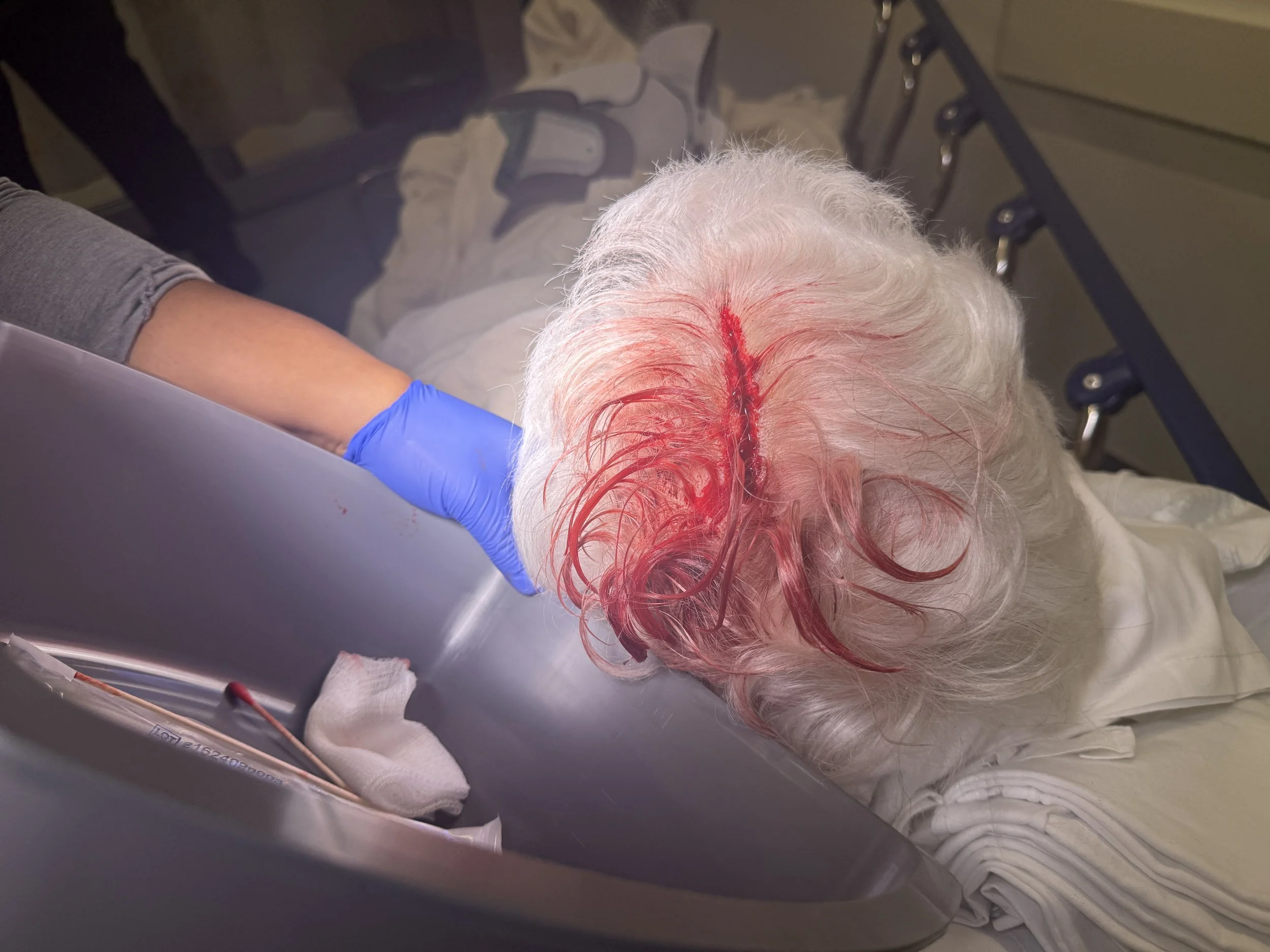
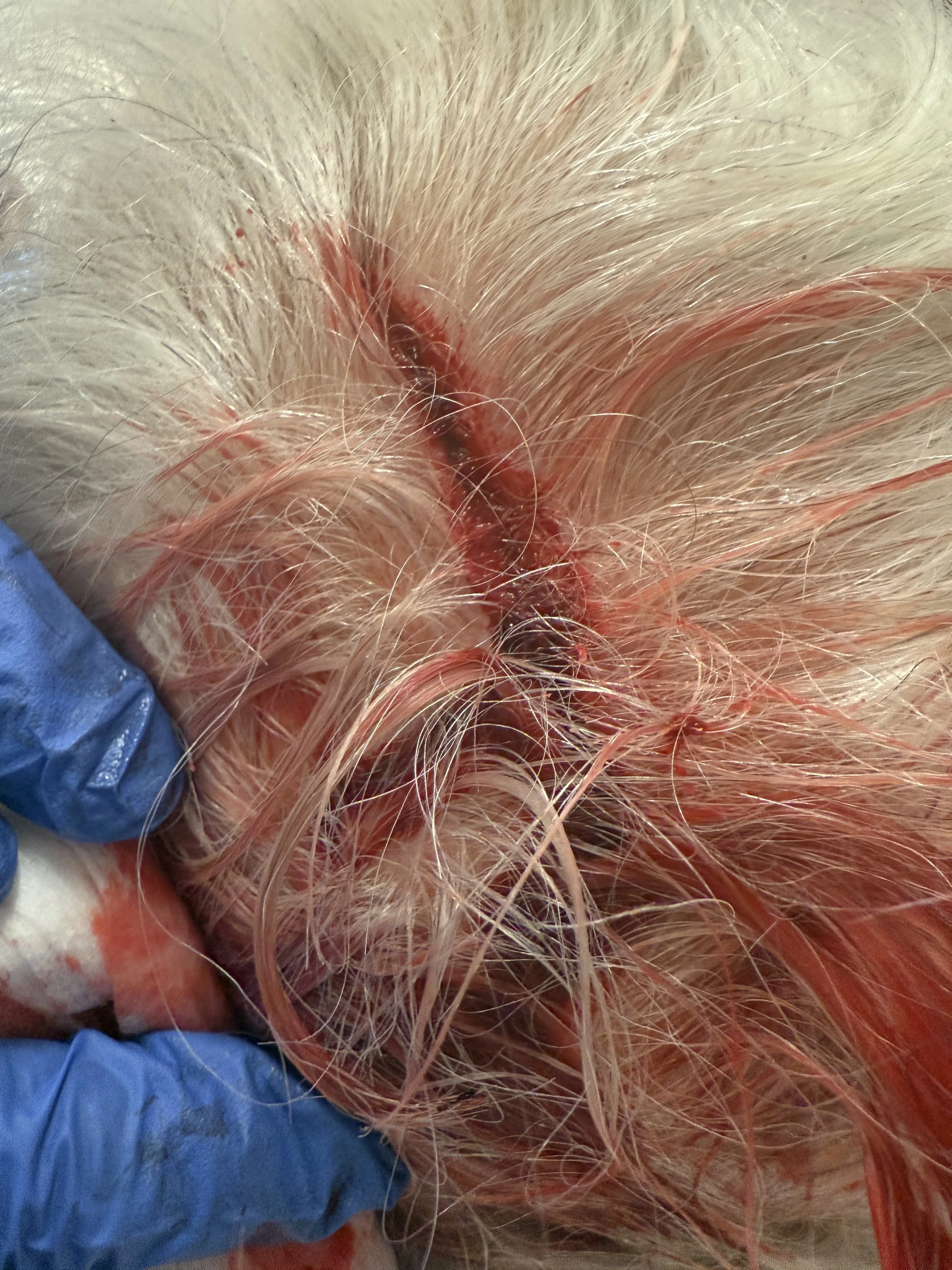
Hemostasis first: Use direct pressure, lidocaine with epinephrine infiltration, or topical hemostatic agents if needed. The scalp is highly vascular, and control of bleeding improves visualization.
Hair management: Part hair with sterile water or gel; shaving is not required and may increase infection risk.
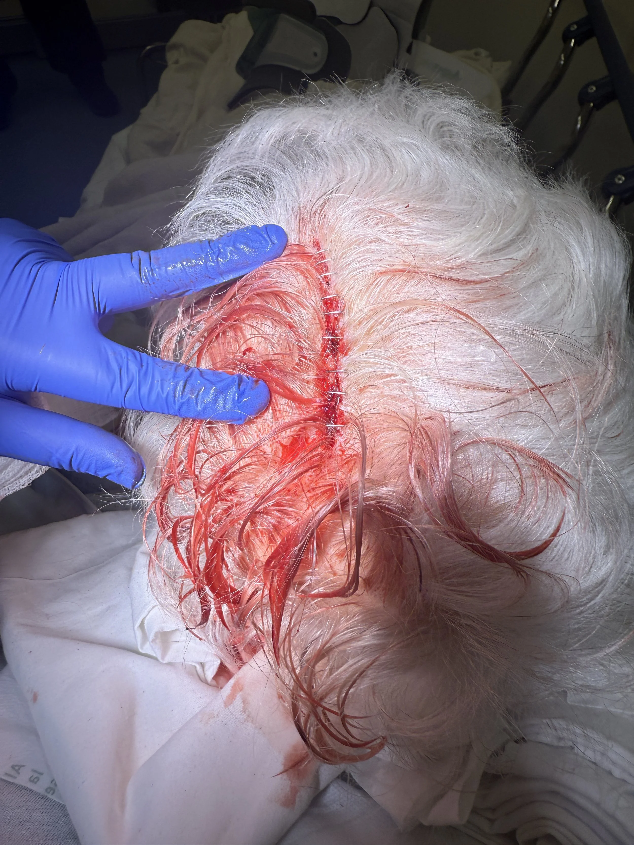
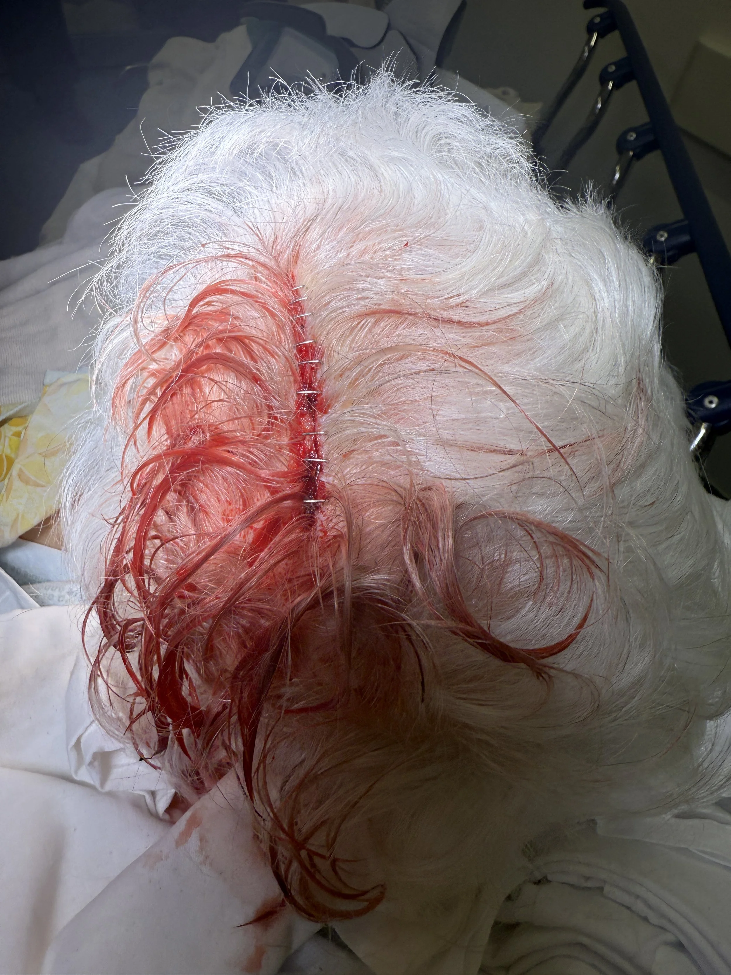
Approximation: Evert edges gently with a skin hook or gloved fingers. The staple should approximate the epidermis without burying deep into dermis or galea. Repairing the galea is important if it is involved - and a much longer process. You will need to use dis- solvable sutures for this and be sure to approximate well.
Staple angle: Place perpendicular to the wound, ensuring even distribution across the wound line. Overlapping staples or angled placement risks poor alignment. You will see a blue indicator on the staple gun - keep this in the middle of the wound when adding staples.
Spacing: Typically 5–10 mm apart, depending on wound tension.
When not to staple:
As mentioned - Galeal lacerations >0.5 cm require layered repair with absorbable sutures before stapling.
Complex, jagged wounds may benefit from sutures for precise approximation.
Post-Procedure Wound Care
Staples may be simple, but wound care is essential for good outcomes:
Dressings: Scalp wounds generally don’t require occlusive dressings once hemostasis is achieved. A light, non-adherent dressing or gauze can be applied for 24 hours if oozing persists.
Cleansing: Patients may gently wash hair with mild shampoo after 24–48 hours. Avoid scrubbing or hair dye products until staples are removed.
Staple removal timing:
7–10 days for most scalp wounds. Some possibly up to 11 or 12 days.
Longer if there is delayed healing (common in elderly or patients with vascular disease).
Infection risk: Relatively low in the scalp due to excellent vascular supply, but instruct patients to return if they develop redness, discharge, or spreading tenderness.
The Emergency Department Checklist
When faced with an elderly patient with a bleeding scalp laceration:
Stabilize and assess: Airway, bleeding control, neurologic exam.
Decide on imaging: In patients ≥65 years, err on the side of head CT. Anticoagulation tips the balance strongly toward imaging.
Repair efficiently: Staples are fast, effective, and cosmetically sound for most scalp wounds.
Layer when needed: Deep galeal injuries require absorbable sutures beneath staples.
Educate: Provide clear wound care instructions, highlight signs of infection, and set follow-up for staple removal.
The Pause
The procedural pause here reminds us: a scalp laceration in the elderly is rarely “just a lac.” It’s an opportunity to catch an intracranial hemorrhage, to respect tissue layers, and to use a technique that balances speed with outcome.
Sometimes the pause is less about the closure itself, and more about remembering what lies beneath—both the vascular galea and the vulnerable brain.
watch
References
Roberts & Hedges: Clinical Procedures in Emergency Medicine and Acute Care, Elsevier, 7th edition.
Singer, A. J., Hollander, J. E., Valentine, S. M., & Turque, T. W. (2002). Prospective, randomized, controlled trial of wound closure with staples versus sutures: cosmetic results and complication rates. Annals of Emergency Medicine, 39(1), 6–13.
Stiell, I. G., Wells, G. A., Vandemheen, K., Clement, C., Lesiuk, H., Laupacis, A., & McKnight, R. D. (2001). The Canadian CT Head Rule for patients with minor head injury. Lancet, 357(9266), 1391–1396.
Haydel, M. J., Preston, C. A., Mills, T. J., Luber, S., Blaudeau, E., & DeBlieux, P. M. (2000). Indications for computed tomography in patients with minor head injury. New England Journal of Medicine, 343(2), 100–105.