Flap Closure for Lower Extremity Wounds and Suture Techniques
Lower extremity lacerations present a unique set of challenges in the emergency department. The skin over the shin, calf, ankle, and foot is notoriously tight, with little redundancy. Vascular supply is less robust than in the face or scalp, and these wounds often exist in the context of trauma, contamination, or high shear forces. Primary closure using simple interrupted sutures may work for straightforward wounds, but when approximation creates excessive tension—or worse, when wound edges blanch—a different approach is needed.
This is where flap closure comes into play. By recruiting local tissue and redistributing tension, flap techniques allow for wound closure that respects blood supply and promotes better healing.

Why Consider a Flap in the Lower Extremity?
Unlike the scalp or face, lower extremity wounds often resist approximation due to the combination of taut skin, minimal underlying soft tissue, and frequent contamination. Attempting to simply “pull the edges together” can create ischemia, wound edge necrosis, or dehiscence. Flaps provide:
Redistribution of tension away from fragile wound edges.
Preservation of vascular supply by moving tissue rather than stretching it.
Improved alignment with relaxed skin tension lines, which optimizes cosmetic outcome and function.
Plastic surgery and trauma literature consistently emphasize that vascularity is king. Flap survival hinges not on the strength of your knots, but on whether the flap’s microcirculation remains intact (Janis & Kwon, 2016).
Technique: Mattress Sutures to Protect the Flap
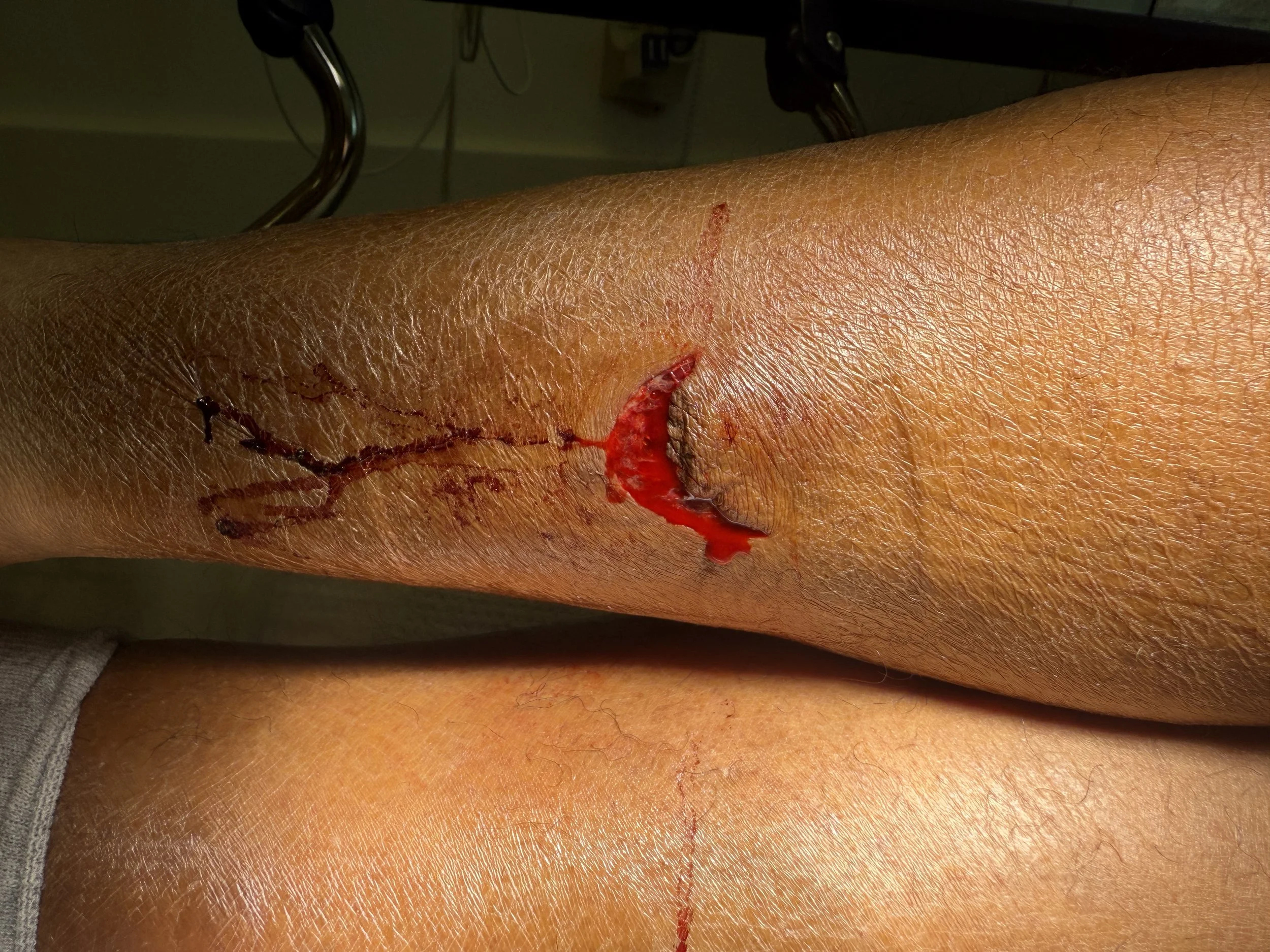
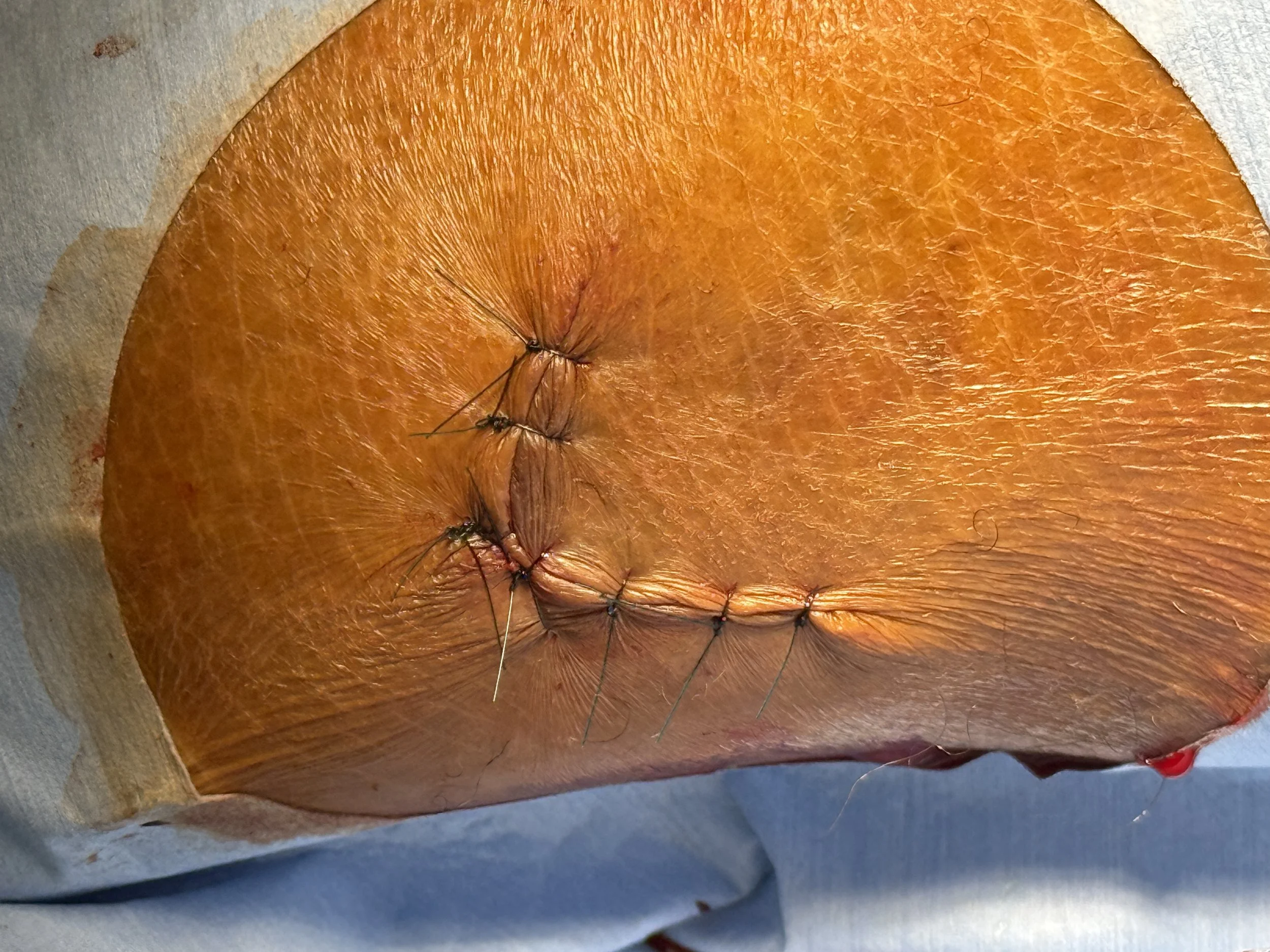
In the case shown here, a V-shaped flap laceration of the lower extremity was closed using a combination of vertical mattress sutures across the flap and simple interrupted sutures for the remaining wound.
Vertical mattress sutures: Ideal for areas of high tension. They provide eversion of wound edges, distribute force deeper in the dermis, and reduce the risk of inversion that compromises vascularity. They also relieve pressure from the fragile flap tip.
Simple interrupted sutures: Still the backbone of laceration repair. They allow for precise edge approximation and are forgiving if infection develops, since a single suture can be removed without compromising the entire closure.
Pearl: Always assess the flap tip for perfusion after placement of tension-relieving sutures. If blanching persists, consider loosening or revising your approach. Overzealous tension can strangle the very tissue you are trying to preserve.
Irrigation and the Challenge of Puncture Wounds
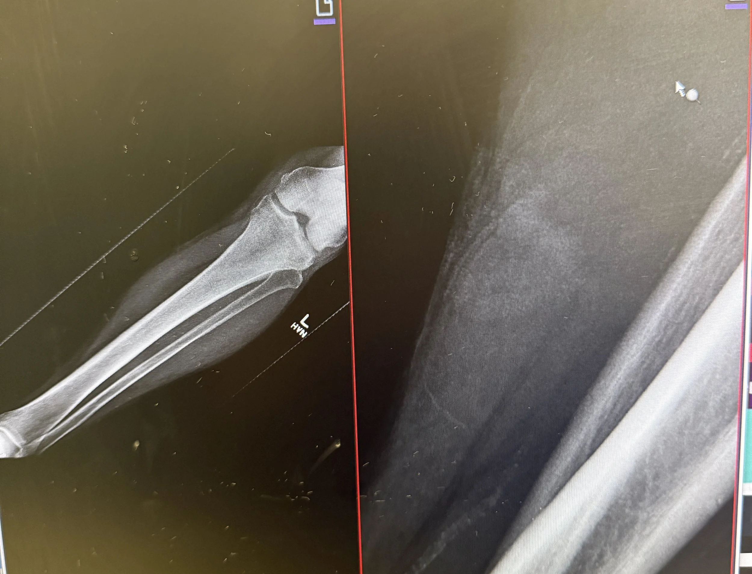
Before closing any wound—especially on the lower extremity—the foundation is irrigation. Contaminated wounds, puncture injuries, and bites carry significantly higher infection risks.
Irrigation Evidence
Volume matters more than pressure. The classic teaching is “the solution to pollution is dilution.” A Cochrane review (Fernandez & Griffiths, 2012) suggests irrigation with normal saline reduces infection risk, but the exact solution is less important than the volume delivered.
Pressure irrigation (7–8 psi), typically achieved with a 19-gauge catheter and a 35–50 mL syringe, has been shown to effectively reduce bacterial load without damaging tissue (Singer & Dagum, 2008).
Tap water vs. saline: Several randomized controlled trials demonstrate no significant difference in infection outcomes between tap water and sterile saline for wound irrigation (Quinn et al., 2014). For lower extremity wounds with heavy contamination, generous irrigation with either is acceptable.
Puncture Wounds
Puncture wounds of the foot or leg are particularly treacherous. Unlike linear lacerations, punctures drive bacteria deep into tissue, often inoculating bone or joint spaces.
Infection risk: Approximately 10% of puncture wounds become infected, and 1–2% progress to osteomyelitis (Cummings & Del Beccaro, 1992). Pseudomonas is classically associated with plantar punctures through athletic shoes.
Irrigation caveat: Aggressive high-pressure irrigation of puncture wounds is controversial. Some evidence suggests it may drive bacteria deeper (Fleisher & Ludwig, 2010). Instead, punctures should be gently irrigated with copious low-pressure solution to reduce superficial contamination without forcing pathogens into deeper structures.
Closure: Most puncture wounds are not closed primarily, especially when contaminated or involving the sole of the foot. Leaving them open, or at most loosely approximated, allows for drainage and reduces infection risk.
Antibiotics: Prophylaxis remains debated, but is often considered for plantar punctures, delayed presentation (>6 hours), or in immunocompromised patients. Coverage should target Staphylococcus aureus and Pseudomonas when shoes are involved.
Aftercare and Healing in the Lower Extremity
Even with meticulous closure, lower extremity wounds are prone to swelling, dehiscence, and delayed healing. Strategies to improve outcomes include:
Immobilization: Splinting or limiting motion across the wound site reduces shear.
Elevation: Encourages venous return and reduces dependent edema that stresses sutures.
Dressing selection: Non-adherent dressings with light compression can protect the wound while minimizing tension.
Patient education: Discuss the slower healing trajectory of leg wounds compared to the face or scalp, and emphasize return precautions for infection, flap necrosis, or wound separation.
The Procedural Pause
Before you tie your first knot, pause. Consider:
Is this wound under excessive tension that will compromise vascularity?
Should I create a flap to redistribute force and protect the tissue tip?
Am I irrigating thoroughly and tailoring my approach for laceration versus puncture?
Does this wound demand a closure at all, or is healing by secondary intention safer?
Every wound is different, but the principles remain constant: respect tissue, protect blood supply, minimize tension, and prevent infection.
Flap Closure may take a combination of sutures.
Modified or vertical mattress are great for tension relief.
Consider adding simple interrupted sutures to bring It all together.
Bandage with anti-bacterial ointment and a non-adhesive dressing with suture removal in 10-12 days.
Watch
References
Cummings, P., & Del Beccaro, M. A. (1992). Antibiotics to prevent infection of simple wounds: a meta-analysis of randomized studies. American Journal of Emergency Medicine, 10(5), 347–351.
Fernandez, R., & Griffiths, R. (2012). Water for wound cleansing. Cochrane Database of Systematic Reviews, (2).
Fleisher, G. R., & Ludwig, S. (2010). Textbook of Pediatric Emergency Medicine (6th ed.). Lippincott Williams & Wilkins.
Janis, J. E., & Kwon, R. K. (2016). Understanding the basic anatomy and physiology of skin flaps. Seminars in Plastic Surgery, 30(1), 3–11.
Quinn, J., Cummings, S., Callaham, M., & Sellers, K. (2014). Suturing versus conservative management of lacerations. Cochrane Database of Systematic Reviews, (3).
Roberts & Hedges: Clinical Procedures in Emergency Medicine and Acute Care, Elsevier, 7th edition.
Singer, A. J., & Dagum, A. B. (2008). Current management of acute cutaneous wounds. New England Journal of Medicine, 359(10), 1037–1046.