The Saline Load Test
A saline load test (SLT) is the most common, non-surgical approach and diagnostic test for traumatic knee injuries involving the joint. The clinician uses a sterile technique to inject saline into the knee (or other joint space) using an 18g needle and syringe (Nord, et. al., 2009). Saline is slowly injected into the joint space until the capsule distends and fluid leeks out the wound, proving for a positive test. Saline is then aspirated back from the knee to remove the pressure. Positive tests will result in a patient being taken to the operating room for wash out and repair.

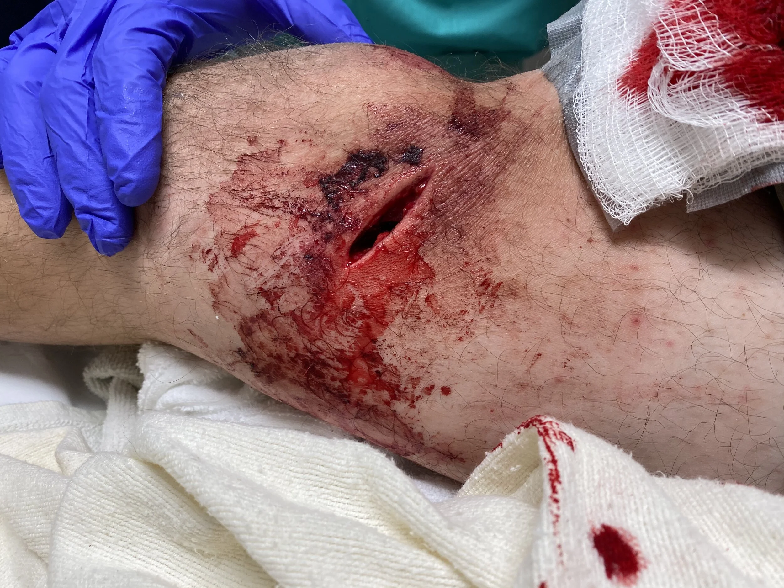
Image: Performing a saline load testing on the right knee, with suspicion of violation of the joint space, from penetrating trauma. Image: M. Roberts, NP.
Joint Arthrography and Saline Load Testing (SLT) - It’s Still a Useful and Accurate Procedure for Penetrating Trauma to the Knee.

The Case
A 30-year-old male arrives to your fast track for a “knee injury”. He was moving boxes and a metal rod poked him and caused a “gash” to the knee. He had no other trauma. You remove the bandages to reveal a small, but deep laceration to the right medial surface to the knee. It’s bleeding and difficult to explore. When you take a closer look, you are concerned the wound penetrates the joint. You obtain radiographs that shows questionable air in the joint and this leads you to further conclude the joint space may have been traumatically violated. Further investigation may be warranted. You have to decide between getting additional imaging, such as a CT or completing a saline load test to confirm your diagnosis. Note: If there is obvious air on the image, immediate consultation with the orthopedic clinician and OR wash out is then indicated without further testing. A CT may be required to assess for further injuries, depending on mechanism of injury and clinical circumstances.
Note: Before saline was used in the SLT, methylene blue was injected into the joint space to reveal joint violation from open wounds to the joint space. But, methalene blue will interfere with arthroscopic evaluation and produce inflammatory reactions. Saline is now used to infiltrate the joint space to confirm diagnosis and is preferred (Roberts & Hedges, 2019).
Time to Proceed
- Ensure your patient is supine and pre-medicated prior to the procedure. We suggest intravenous pain medication, as these injuries can be extremely painful. The injection causes pressure and pain.
- Good lighting is a must.
- Ask a colleague to assist with the procedure.
- Obtain two-three 50 mL syringes and an 18 g. or 20 g. needle as well as sterile saline and Chlorhexidine wash, sterile gloves and drapes.
- Aseptic technique is essential but the equipment and procedure are the same as performing arthrocentesis with just a few differences.
- You may consider a set up using a sterile saline bag of fluid and a stopcock to control the flow as you do the test. We suggest a more focused and slow approach using syringes.
- The goal is to “load” the joint space with the saline as you slowly inject it into the joint space.
- You should position yourself on the opposite side of injury for the injection.
- You must inject 50-200 mL of sterile saline (for an adult patient) into the knee to sufficiently create enough volume in the joint to visibly distend the joint and cause leakage from a violated joint space out into the surrounding tissues.
- After you confirm or deny a diagnosis, remove the saline from the joint space by aspirating back the fluid into the syringe.
- Elevate the knee post injection and apply ice packs to assist with comfort.
- Ensure bleeding is controlled by applying pressure to the area.
Positive test
A positive test for joint compromise from a penetrating injury will reveal leakage of fluid from the violated joint space into the surrounding tissue/wound, usually dripping out slowly as you finish injecting the saline into the joint space.
Negative test
A negative test will reveal the opposite; there will be an absence of evidence of leakage after the appropriate amount of saline is injected into the joint space.

Image above: right knee, with medial laceration. An example of a positive SLT, as the fluid from the saline injected into the joint space from the opposing side causes fluid to leak out from the violated joint space. Image by: M. Roberts, NP.
Check out a real case and watch the STEP-BY-STEP video showing you how to perform SLT.
Click the video below!
SLT Pearls
You may consider using SLT for other joints such as the ankle or wrist. These smaller joints require much less saline. You may also consider using ultrasound to confirm your placement and volume of injection into the space.
Knee: 100-200 mL of saline
Elbow or Ankle: 20-30 of saline
Wrist: 5 mL of saline
Shoulder: 40-60 mL of saline of saline (Roberts & Hedges, 2019).
When uncertain about the plan or the procedure, discuss it with the orthopedic specialist/consultant.
Time to Process
Radiograph + SLT +/- a CT?
There are orthopedic specialists and emergency department clinicians who believe that focused imaging, such as a CT may be beneficial for the patient either to replace the SLT or to use in addition to the SLT. You should discuss this with the orthopedic clinician on call in your department and share with them the case and radiographs. Should they prefer to use SLT prior to advanced imaging, you can either assist with the procedure or perform it before the orthopedic consultant is able to arrive to the department to see the patient. Patients with a positive SLT test should be taken to the OR for wash out and other potential procedures.
The SLT can be extremely accurate. But, if you do not inject enough saline into the joint, you may decrease your sensitivity. To achieve 95% sensitivity of the SLT into the knee, you must insert at least 194 mLs of saline into the joint (Keese et. al, 2007). In addition a saline solution load volume of 47 mL is required to detect 90% of superolateral traumatic arthrotomies of 5 mm in the pediatric knee with use of the SLT . But, some clinicians suggest not to use SLT alone to confirm the diagnosis. Plain film radiographs and expert consultation for CT imaging is imperative. CT imaging will show a more definitive presence of air in the joint space if questionable on the radiograph. Patients with true air in the joint on the CT are taken to the OR, while patients with negative CTs are sent home from the ER if no indications for further testing or admission is present. Therefore, a CT may prevent the patient from needing additional hospital resources. These are all things to take into consideration when deciding on formal imaging and disposition.
In the end, the sensitivity and specificity of a CT scan to detect traumatic arthrotomies can be as accurate as 100%. Finally, additional benefits of obtaining a CT scan are that it can improve detection and treatment of open periarticular knee fractures as compared to using plain radiograph to detect a fracture, which can be as low as 65% (Konda, et al., 2013).
References
Haller JM, Beckmann JT, Kapron AL, et al. Detection of a traumatic arthrotomy in the pediatric knee using the saline solution load test. Journal of Bone and Joint Surgery 2015;97(10):846-849.
Keese, G, Boody, A, Wongworwat, M, Jobe, C. The accuracy of the saline laod test in the diagnosis of traumatic knee arthrotomies. Journal of Orthopaedic Trauma. Aug; 2007. 21(7): 442-433.
Konda S, Howard D, Davidovitch R, Egol K. The role of computed tomography in the assessment of open periarticular fractures associated with deep knee wounds. Journal of Orthopaedic Trauma. 2013 Sep; 27(9):509-14
Nord RM, Quach T, Walsh M, et al. Detection of traumatic arthrotomy of the knee using the saline solution load test. Journal of Bone and Joint Surgery. 2009;91(1):66-70.
Roberts JR, Custalow C, Hedges JR, et al. Roberts and Hedges’ Clinical Procedures in Emergency Medicine and Acute Care. 7th ed. Philadelphia, PA: Elsevier; 2013.1092:4.